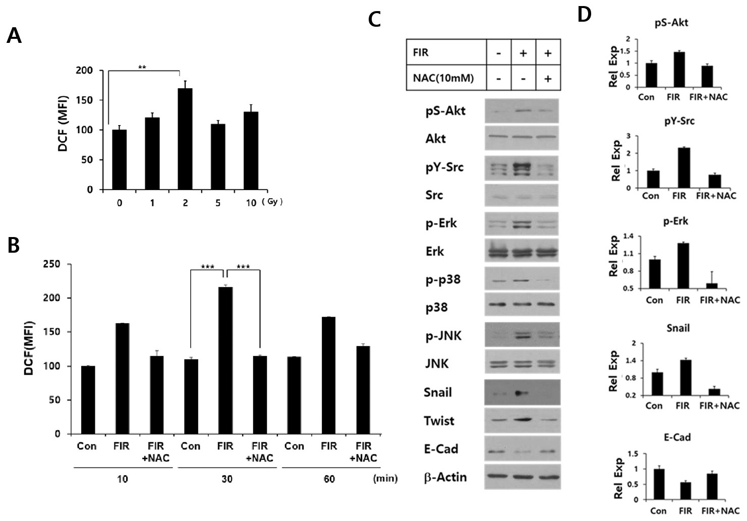
Fig. 1. FIR up-regulates intracellular ROS which mediates EMT signaling by FIR in colorectal cancer cells. (A) HCT116 p53+/+cells were irradiated with indicated doses using the gamma irradiator, and the intracellular ROS levels were determined at 30 min by H2DCFDA staining as described in the text. (B) HCT116 p53 +/+ cells received fractionated irradiation (FIR) administered at 2 Gy per day for three consecutive days. NAC pretreatment was done 1 h prior to the last irradiation and the ROS levels were determined at indicated time points after irradiation. Results show data (mean + SE) obtained from 3 experiments performed in triplicate wells (**, p ≤ 0.01; ***, p ≤ 0.005). (C) Cells received FIR as described in panel B (2 Gy for 3 days) were incubated for 2 h after the last irradiation, and subjected to Western blot to analyze EMT markers and signaling molecules. Results show representative blot data obtained from multiple experiments. (D) Densitometry analysis of immunoblots was performed to show the relative expression (Rel Exp) of pS-Akt, pY-Src, and pS/Y-Erk levels normalized to Akt, Src, and Erk levels, respectively. For example, the ratio of pS-Akt over Akt levels in control cells was taken as 1. Likewise, E-cad and Snail expression levels were normalized to beta-actin, and the expression ratio for each protein over beta-actin in the untreated control was taken as 1. Results show data (mean + SE) obtained from 3 experiments.